Focusing on algal-derived phytoexosomes *1 obtained from marine algae that survive in harsh environments, we confirmed multiple effects related to inflammation and cellular aging
, and also investigated their use in combination with vitamin C and vitamin C derivatives.
November 17, 2026
ROHTO Pharmaceutical Co., Ltd. (Headquarters: Osaka City, President: Hidetoshi Segi) is engaged in research on "phytoexosomes" *2, which are exosome-like extracellular vesicles derived from plants, microorganisms, algae, etc.
We investigated the effects of phytoexosomes derived from the marine microalga Pavlova sp., which inhabits Okinawa, on inflammation and cellular senescence using cultured human skin cells. In epidermal keratinocytes *3, we confirmed a decrease in the expression of inflammation-related genes and an increase in the expression of differentiation and barrier formation-related genes. In dermal fibroblasts *4, the increase in aging-related indicators associated with subculturing was suppressed. Furthermore, in epidermal keratinocytes irradiated with ultraviolet light, the combined use of algae-derived phytoexosomes and vitamin C reduced the production of the inflammation-related substance PGE2 *5 compared to each condition alone. In addition, in dermal fibroblasts, the combined use of algae-derived phytoexosomes and a vitamin C derivative increased the formation of type I collagen fibers compared to the use of the derivative alone. These results have been presented at domestic and international academic conferences, including the International Federation of Societies of Cosmetic Chemists (IFSCC) 2025.
Key points of the research
- Comprehensive analysis of epidermal keratinocytes revealed decreased expression of inflammation-related genes and increased expression of differentiation and barrier formation-related genes.
- PGE2 levels, which increased due to UV radiation, decreased with the addition of PGE2 alone, and showed lower values when combined with vitamin C than under each individual condition.
- In a passage aging study of dermal fibroblasts, aging-related indicators such as SA-β-gal, p16, and p21 improved.
- Under conditions of use with a vitamin C derivative, the formation of type I collagen fibers increased compared to when the derivative was used alone.
Research Background
Focusing on the potential of algae to survive in harsh environments, we have developed algae-derived phytoexosomes.
Pavlova sp. is a marine microalga that inhabits the mangrove areas of northern Okinawa Island. It grows while being exposed to various environmental stresses such as drought, ultraviolet radiation, heat, and changes in osmotic pressure. It also contains components that are involved in adaptation to the environment, such as fatty acids like EPA and DHA, fucoxanthin, and betaine lipids.
Therefore, we hypothesized that the vesicles obtained from Pavlova, which survives in such environments, might also possess characteristics related to stress response. To clarify how these properties relate to human skin cells, we focused on inflammation and cellular aging caused by ultraviolet radiation and conducted research using epidermal keratinocytes and dermal fibroblasts.
Multifaceted evaluation of the properties and stability of algae-derived phytoexosomes.
To effectively utilize new materials in skincare, it is crucial to accurately understand not only their functionality but also the properties of the material itself, evaluating its quality, stability, and safety. At our company, we comprehensively evaluate materials by examining not only raw material selection and manufacturing conditions, but also characteristics such as particle size and structure. For the algae-derived phytoexosomes in this study, we confirmed they are lipid bilayer vesicles with an average particle diameter of approximately 160 nm. Furthermore, with a view to product application, we evaluated their stability in emulsified formulations. Algae-derived phytoexosomes were blended into an emulsion base and stored at a high temperature of 60°C for three weeks. The number of particles, particle size, and the retention rate of encapsulated RNA *6 were measured. The results showed that even after three weeks, the number of particles remained at approximately 60% of the initial value, and no significant changes were observed in particle size. Additionally, approximately 70% of the encapsulated RNA was confirmed to remain. These results suggest that algae-derived phytoexosomes exhibit a certain level of stability even under harsh high-temperature conditions in emulsified formulations, indicating their potential for product application (Figure 1).
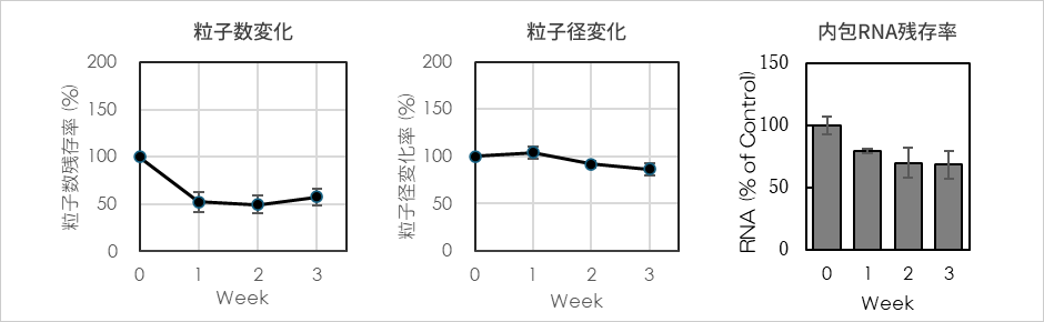
Figure 1: Changes in particle number, particle size, and encapsulated RNA retention rate of algae-derived phytoexosomes formulated in an emulsion substrate.
Furthermore, when algae-derived phytoexosomes were added to cultured human skin cells, we confirmed that they were taken up into epidermal keratinocytes and dermal fibroblasts (Figure 2).
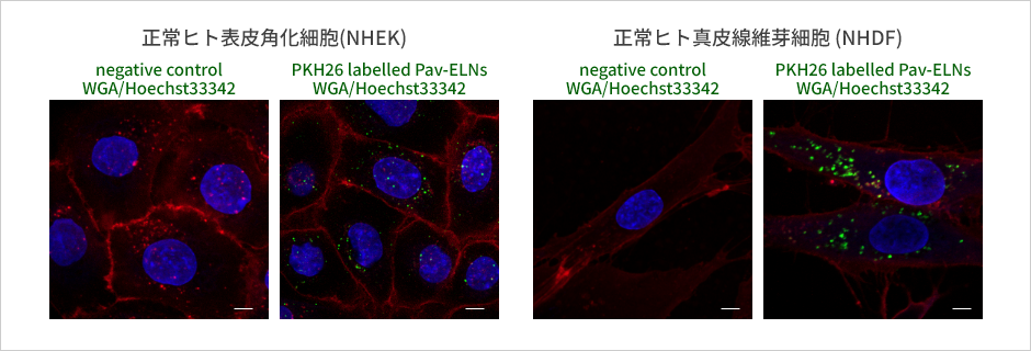
Scale bars: 5µm
Figure 2: How algae-derived phytoexosomes are taken up by cultured human skin cells (green: algae-derived phytoexosomes, red: cell membrane, blue: nucleus).
*The stability results are based on the above test formulation and storage conditions. Furthermore, the evaluation of cell uptake was conducted through an addition test to cultured cells and does not indicate skin penetration of the applied product.
In this study, we evaluated gene expression and cellular responses related to inflammation, differentiation/barrier formation, and cellular senescence, based on the properties of these materials and their behavior in cells. Furthermore, through combination tests with vitamin C and vitamin C derivatives, we investigated not only the effects of algae-derived phytoexosomes themselves, but also their potential when combined with existing skincare ingredients.
result
Result 1: Evaluated the effects of algae-derived phytoexosomes on gene expression in epidermal and dermal cells.
Human skin cells were treated with algae-derived phytoexosomes, and comprehensive gene expression analysis was performed using RNA-seq *7. As a result, gene expression related to inflammation decreased in the epidermis, while gene expression related to epidermal differentiation and barrier formation increased. In the dermis, gene expression related to inflammation and aging decreased, gene expression related to the breakdown of dermal fibers such as collagen and elastin decreased, and gene expression related to fiber formation increased.
These results indicate that changes occurred not only in a single gene, but also in multiple cellular functions, including inflammatory responses, differentiation and barrier formation, and aging suppression.
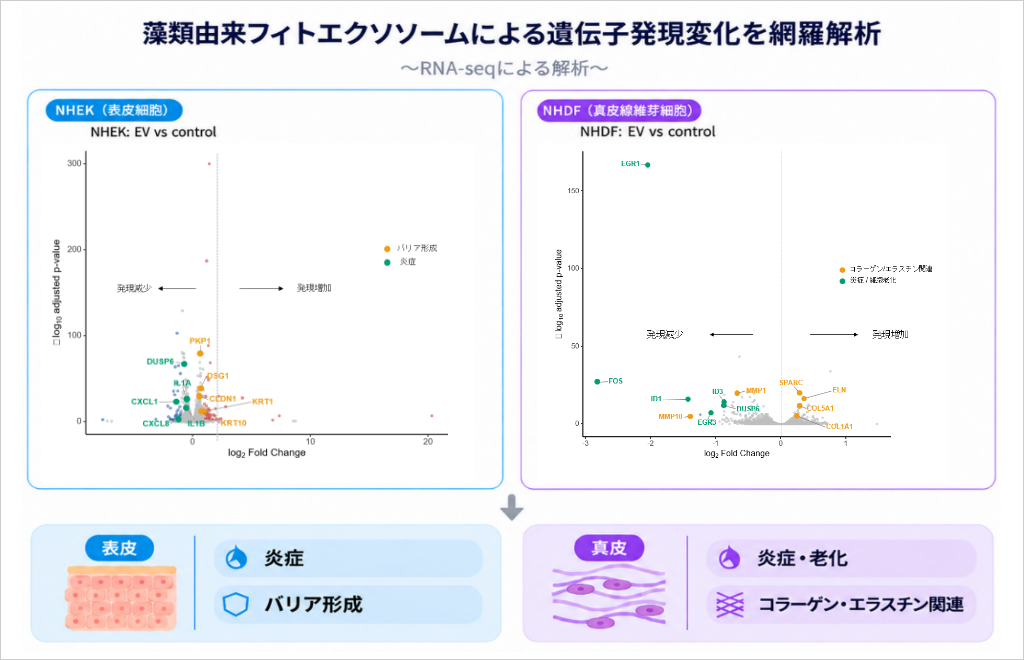
Figure 3: Comprehensive analysis of gene expression changes induced by algae-derived phytoexosomes.
<Testing Method>
Normal human epidermal keratinocytes (NHEK) and normal human dermal fibroblasts (NHDF) were used to compare a group treated with algae-derived phytoexosomes with a control group. RNA was extracted from the cells of each group, and comprehensive gene expression analysis was performed using RNA sequencing (RNA-seq). (Conducted by Rohto Pharmaceutical Research Institute)
Result 2: Confirmed that UV-induced inflammation of epidermal cells is suppressed, and that this is further suppressed when used in combination with vitamin C.
Epidermal cells exposed to ultraviolet (UV) light release inflammatory substances such as IL-6 and prostaglandin E2 (PGE2). These are thought to be involved in redness and skin irritation after sun exposure. When algae-derived phytoexosomes were added to epidermal keratinocytes immediately after irradiation with UVB light and cultured for 72 hours, the secretion of both IL-6 and PGE2, which had increased due to UV light, was suppressed. In addition, while vitamin C alone reduced PGE2 production, when vitamin C was added in combination with algae-derived phytoexosomes, the values were lower than when each substance was added individually (Figure 4).
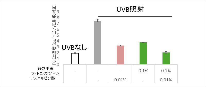
Figure 4: Suppression of UV-induced inflammatory substances by algae-derived phytoexosomes and vitamin C.
<Testing Method>
Normal human epidermal keratinocytes were seeded and irradiated with UVB (25 mJ/cm2) 24 hours later. Algal-derived phytoexosomes and vitamin C were added immediately afterward. After 72 hours of incubation, the culture supernatant was collected, and PGE2 was quantified by ELISA. (n=3, Dunnet, ***P<0.001) (Conducted by Rohto Pharmaceutical Research Institute)
Result 3: The progression of serial aging was suppressed in dermal fibroblasts.
Fibroblasts, which produce collagen and other substances in the dermis, undergo a process called "cellular senescence" as they repeatedly divide, eventually becoming larger and flatter, and ceasing to proliferate. These aged cells are thought to release inflammatory substances into their surroundings, contributing to the aging of the skin.
Therefore, we induced senescence in young fibroblasts by subculturing them more than 20 times in both a medium containing algal-derived phytoexosomes and a medium without them. Under the condition without algal-derived phytoexosomes, the SA-β-gal staining region, a marker of cellular senescence, expanded, and the expression of the senescence-promoting genes p16 and p21 increased. On the other hand, under the condition containing algal-derived phytoexosomes, these changes were suppressed, and no decrease in proliferative capacity was observed (Figure 5).
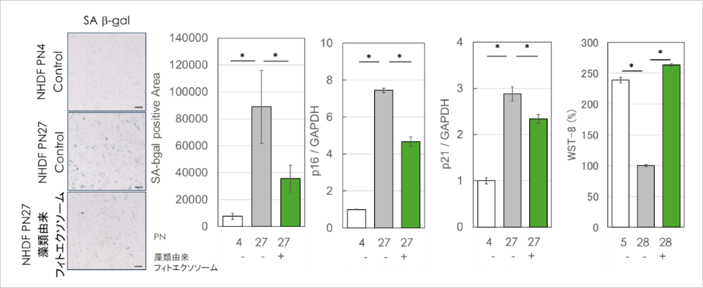
Figure 5: Inhibition of aging progression of dermal fibroblasts by algae-derived phytoexosomes.
<Testing Method>
Normal human dermal fibroblasts at passage 4 were subcultured up to passage 28 (PN) in a medium containing or without algal-derived phytoexosomes. SA-β-gal staining was analyzed by image analysis to quantify positive regions, p16 and p21 gene expression was evaluated by real-time RT-PCR, and proliferation capacity was assessed by WST-8. (n=3 or 4, student's t-test *P<0.05) (Conducted by Rohto Pharmaceutical Research Institute)
Result 4: Confirmed effect on the extracellular matrix that improves the structure of the skin's foundation; increased formation of type I collagen fibers when used in combination with vitamin C derivatives.
The firmness and elasticity of the dermis are supported by fibers such as collagen and elastin (extracellular matrix). When algae-derived phytoexosomes were added to fibroblasts, the gene expression of type I collagen and elastin increased, while the gene expression of MMP1, an enzyme that breaks down collagen, decreased. Vitamin C is also an essential component for collagen synthesis. When algae-derived phytoexosomes were added to fibroblasts cultured with a vitamin C derivative, the formation of type I collagen fibers increased significantly compared to when only the vitamin C derivative was used (Figure 6).
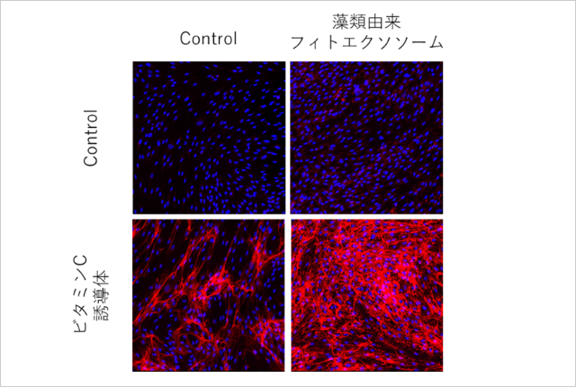
Figure 6: Changes in collagen I fiber formation with the combined use of algae-derived phytoexosomes.
<Testing Method>
Normal human dermal fibroblasts were cultured in a medium containing or without a vitamin C derivative (magnesium ascorbyl phosphate), and algal phytoexosomes were added. Type I collagen fibers were immunofluorescently stained, and positive regions were quantified by image analysis. (Conducted by Rohto Pharmaceutical Research Institute)
Future outlook
In this study, algae-derived phytoexosomes were found to decrease the expression of inflammation-related genes and increase the expression of differentiation and barrier-forming-related genes in epidermal keratinocytes, and to suppress serial aging and promote collagen fiber synthesis in dermal fibroblasts. These results suggest that this material may be involved in multiple cellular functions related to maintaining skin homeostasis. Furthermore, the combination with vitamin C investigated in this study resulted in lower PGE2 production than under each individual condition, and the combination with a vitamin C derivative increased type I collagen fiber formation compared to the derivative alone. In addition to the effects of algae-derived phytoexosomes themselves, it is expected that combining them with existing ingredients may further enhance the value of skincare.
In order to establish phytoexosomes as a new research area, it is important to address various challenges, including elucidating their function, evaluating quality based on differences in origin and manufacturing conditions, developing technologies for stable production, and establishing methods for evaluating transdermal absorption.
Going forward, our company will implement "Longevity" science in skincare, and based on this research, we will advance technological development for material quality evaluation and stable production, aiming to implement the research results in society.
About phytoexosome research
Under the guidance of Professor Takahiro Ochitani of Tokyo Medical University, our company has been promoting phytoexosome research.
Terminology
*1: Algae-derived phytoexosomes: Phytoexosomes derived from the microalga Pavlova sp., which inhabits Okinawa.
*2: Phytoexosomes: Extracellular vesicles of natural origin, such as those found in plants, microorganisms, and algae, excluding animals.
*3: Epidermal keratinocytes: Cells that make up the majority of the "epidermis," the outermost layer of the skin. They are born in the deeper layers of the epidermis and migrate to the surface as they mature, forming the stratum corneum. They play an important role in maintaining the "barrier function" that keeps the skin moist and protects the skin from external stimuli.
*4: Dermal fibroblasts: Cells that produce collagen and elastin in the inner layer of the skin (dermis), supporting the firmness and elasticity of the skin.
*5: PGE2: An abbreviation for prostaglandin E2, a type of physiologically active substance produced in the body that is involved in reactions such as inflammation. It is also produced in the skin when exposed to ultraviolet light and has the function of promoting melanin production.
*6: Encapsulated RNA: RNA contained inside tiny particles such as extracellular vesicles. RNA is a molecule involved in the transmission of genetic information and the regulation of gene function, and when encapsulated within particles, it can be involved in intercellular communication.
*7: RNA-seq: An abbreviation for RNA sequencing, a technique used to investigate which genes are actively functioning and to what extent (gene expression) in specific cells or tissues. This involves reading RNA sequence information to identify and measure the amount of activated genes.